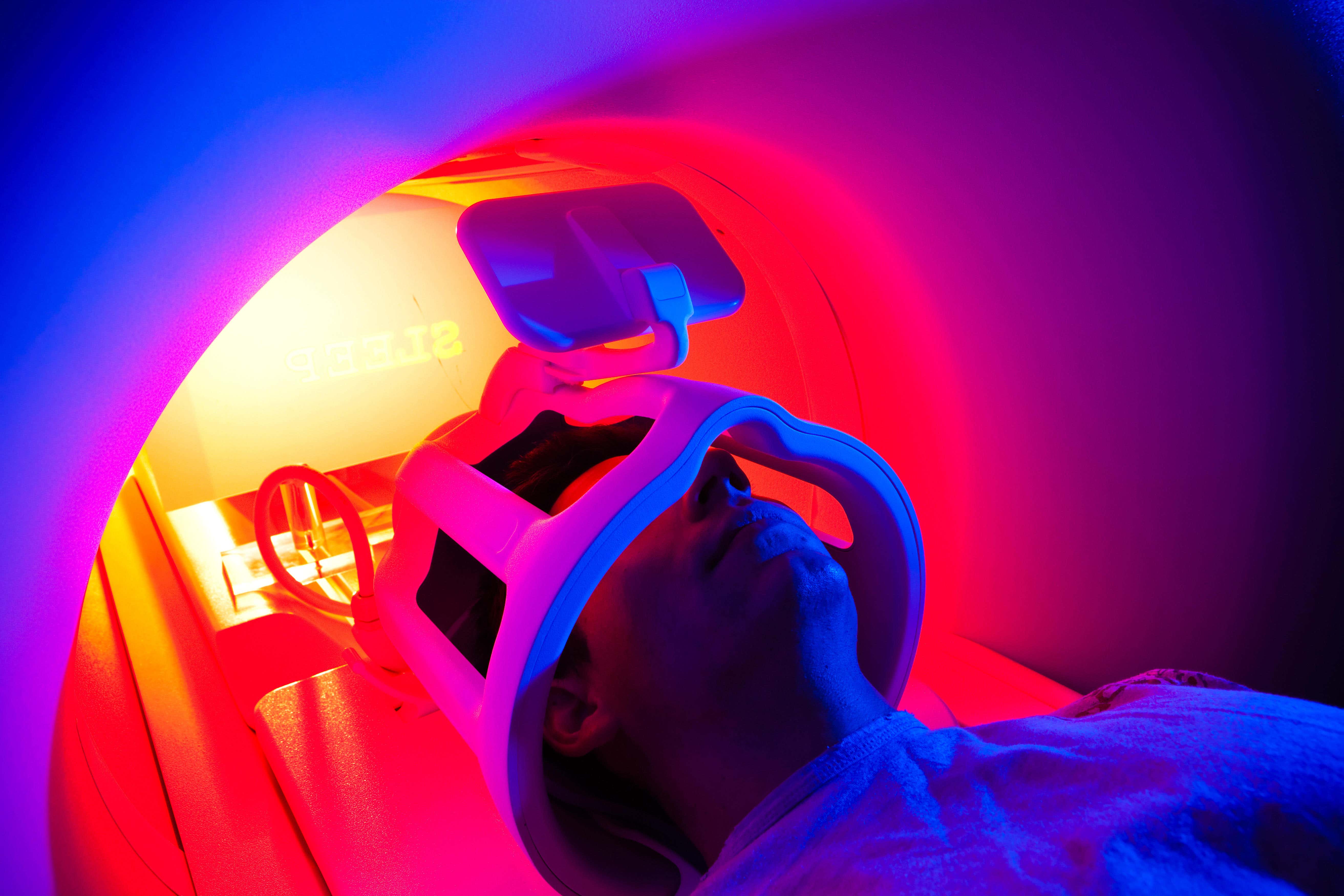
Routine brain scans could pinpoint underlying cause of psychosis, study suggests
Researchers analysed more than 1,600 cases to identify any secondary, physical causes.

Your support helps us to tell the story
From reproductive rights to climate change to Big Tech, The Independent is on the ground when the story is developing. Whether it's investigating the financials of Elon Musk's pro-Trump PAC or producing our latest documentary, 'The A Word', which shines a light on the American women fighting for reproductive rights, we know how important it is to parse out the facts from the messaging.
At such a critical moment in US history, we need reporters on the ground. Your donation allows us to keep sending journalists to speak to both sides of the story.
The Independent is trusted by Americans across the entire political spectrum. And unlike many other quality news outlets, we choose not to lock Americans out of our reporting and analysis with paywalls. We believe quality journalism should be available to everyone, paid for by those who can afford it.
Your support makes all the difference.Offering routine brain scans to people experiencing psychotic episodes could help identify underlying conditions causing their symptoms, a new study has found.
Researchers from the University of Oxford looked at more than 1,600 cases of first-episode psychosis (FEP) – a term used to describe when a person first has a psychotic episode – to identify any secondary, physical causes.
The study – which has been published in the JAMA Psychiatry journal – said “failure to detect such cases” in their early stages “can have serious clinical consequences”, and it has been suggested that MRIs should be “mandatory” for all FEP patients.
However, it added it “remains a controversial issue, partly because the prevalence of clinically relevant MRI abnormalities in this group is unclear”.
Our findings suggest that MRI scans should be considered as part of the initial assessment of all people with first-episode psychosis to ensure that they get the right diagnosis and the right treatment
The research team defines “clinically relevant” abnormalities as an abnormality which led to a change in management, such as a referral to specialists.
They were grouped into the following categories: white matter, vascular, ventricular, cyst, pituitary, tumour, cerebral atrophy, and other.
Twelve independent studies comprising 1,613 FEP patients were analysed, and 26.4% were found to have an intracranial radiological abnormality, while 5.9% had a clinically relevant abnormality.
The most common type were white matter abnormalities (0.9%), followed by cysts (0.5%).
Dr Graham Blackman from the University of Oxford, who led the research team alongside Prof Philip McGuire, said: “Patients presenting with psychosis may have another physical illness or condition causing their symptoms that can be identified using MRI scanning.
“A failure to detect these causes at an early stage can have serious consequences, such as a delay in providing the appropriate treatment.
“Our findings suggest that MRI scans should be considered as part of the initial assessment of all people with first-episode psychosis to ensure that they get the right diagnosis and the right treatment.”
This new evidence has important implications for clinical care in psychosis and a review of the NICE guidance in this area would be helpful
According to the study it is considered good practice to offer a brain scan to new patients with psychosis, but is not mandatory.
A technology appraisal by the National Institute for Health and Care Excellence (Nice), which was published in February 2008 and reviewed in 2011, was unable to recommend scanning as a routine part of the initial investigations into FEP patients.
Prof McGuire added: “We feel that this study addresses a critical knowledge gap in this area by showing that clinically relevant abnormalities occur frequently enough to justify making MRI scanning a routine part of the assessment of people presenting with psychosis for the first time.
“This new evidence has important implications for clinical care in psychosis and a review of the NICE guidance in this area would be helpful.”