Cadavers in the cloud would help doctors treat illness, claim scientists
A detailed 3D copy of your body could let your doctor diagnose illness or even rebuild body parts

Your support helps us to tell the story
From reproductive rights to climate change to Big Tech, The Independent is on the ground when the story is developing. Whether it's investigating the financials of Elon Musk's pro-Trump PAC or producing our latest documentary, 'The A Word', which shines a light on the American women fighting for reproductive rights, we know how important it is to parse out the facts from the messaging.
At such a critical moment in US history, we need reporters on the ground. Your donation allows us to keep sending journalists to speak to both sides of the story.
The Independent is trusted by Americans across the entire political spectrum. And unlike many other quality news outlets, we choose not to lock Americans out of our reporting and analysis with paywalls. We believe quality journalism should be available to everyone, paid for by those who can afford it.
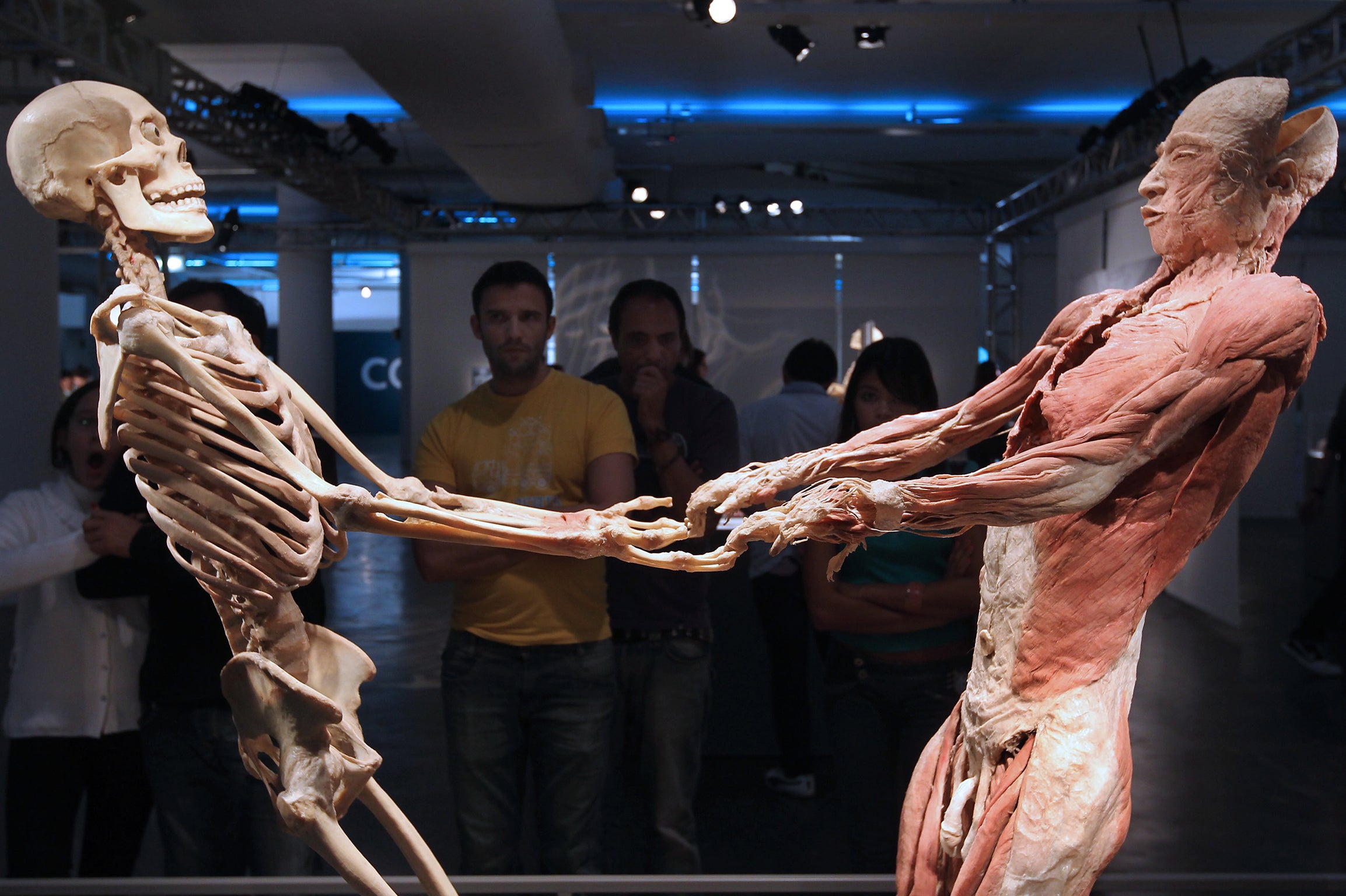
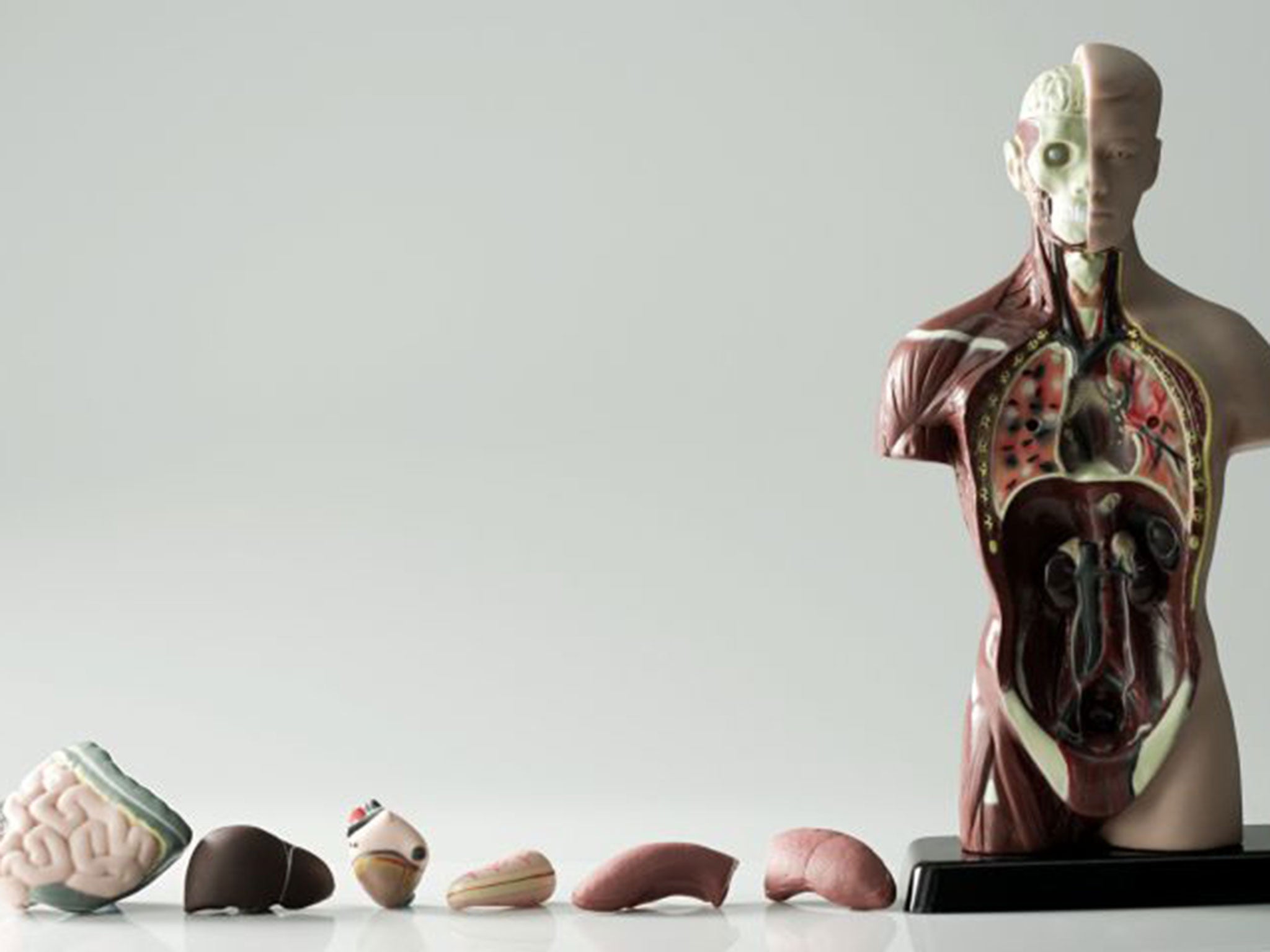
Your support makes all the difference.People could one day have a virtual twin body stored in cyberspace – a cadaver in the cloud – allowing doctors to compare it against the patient’s actual body to help treat illness, scientists have predicted.
The technology to store a high-resolution, three-dimensional copy of body organs and tissues already exists, and James Mah, a surgical imaging specialist at the University of Nevada in Las Vegas, says some medical schools are already using table-top screens to perform training dissections on virtual cadavers.
Dr Mah said the US military was investigating the possibility of storing 3D full-body scans of soldiers so that battlefield surgeons could compare an injured soldier’s body with their healthy cyber-twin stored on the internet.
It may even be possible one day to use full-sized virtual bodies as templates for making replacement body parts, which can be sent over the internet to 3D printers situated near a battlefield.

“The idea is to image somebody in the healthy state so that the data is available if it is needed at a later point. This has some military applications because we have soldiers who get injured and lose limbs and other tissues and it’s a challenge to reconstruct [them],” Dr Mah said.
“The thinking is, if soldiers were imaged beforehand, it might be possible to at least provide a surgical template that could be printed in the field to facilitate surgical repair. That would be a great benefit in the field,” he told the American Association for the Advancement of Science conference in San Jose, California.
The development has come out of the ability to merge digital images and data from a range of hospital scanners, such as X-rays, MRI and CT. By blending the various scans of a patient, a complete three-dimensional picture of the body can be built up and shown on a full-sized screen which can be manipulated as easily as a picture on a phone or tablet.
“It’s meant to be a life-sized person on a table,” he told the meeting. “We take two-dimensional data and process it into a three-dimensional, touch-sensitive display.
“It allows a person to interact with it by touching it. If we want to see through it we can remove the different layers. At any point we can rotate it and zoom in to get deeper into the anatomy.”
Each layer of tissue can be removed, peeled away or sliced by a virtual scalpel. This enables medical students to perform virtual operations on the same body several times without damaging a real donor cadaver.
There are many other applications for the technology, such as storing a patient’s various hospital scans as a virtual body, available in cyberspace for recall when needed by doctors or surgeons.
“Once you have three-dimensional data, the applications are almost endless,” Dr Mah said. “The obvious one is for education. Then there is the clinical use to preview the patient in advance for more detailed planning. With three-dimensional planning they can reduce the number of possible outcomes and so be more efficient in surgery.”
A big advantage for medical schools is in circumventing the shortage of real cadavers. “In terms of education, the access to human cadaver material has diminished over the years due to an increase in regulation and other factors. So, there is a void in medical education where in many countries there are no long cadaver labs,” he said.
Join our commenting forum
Join thought-provoking conversations, follow other Independent readers and see their replies
Comments