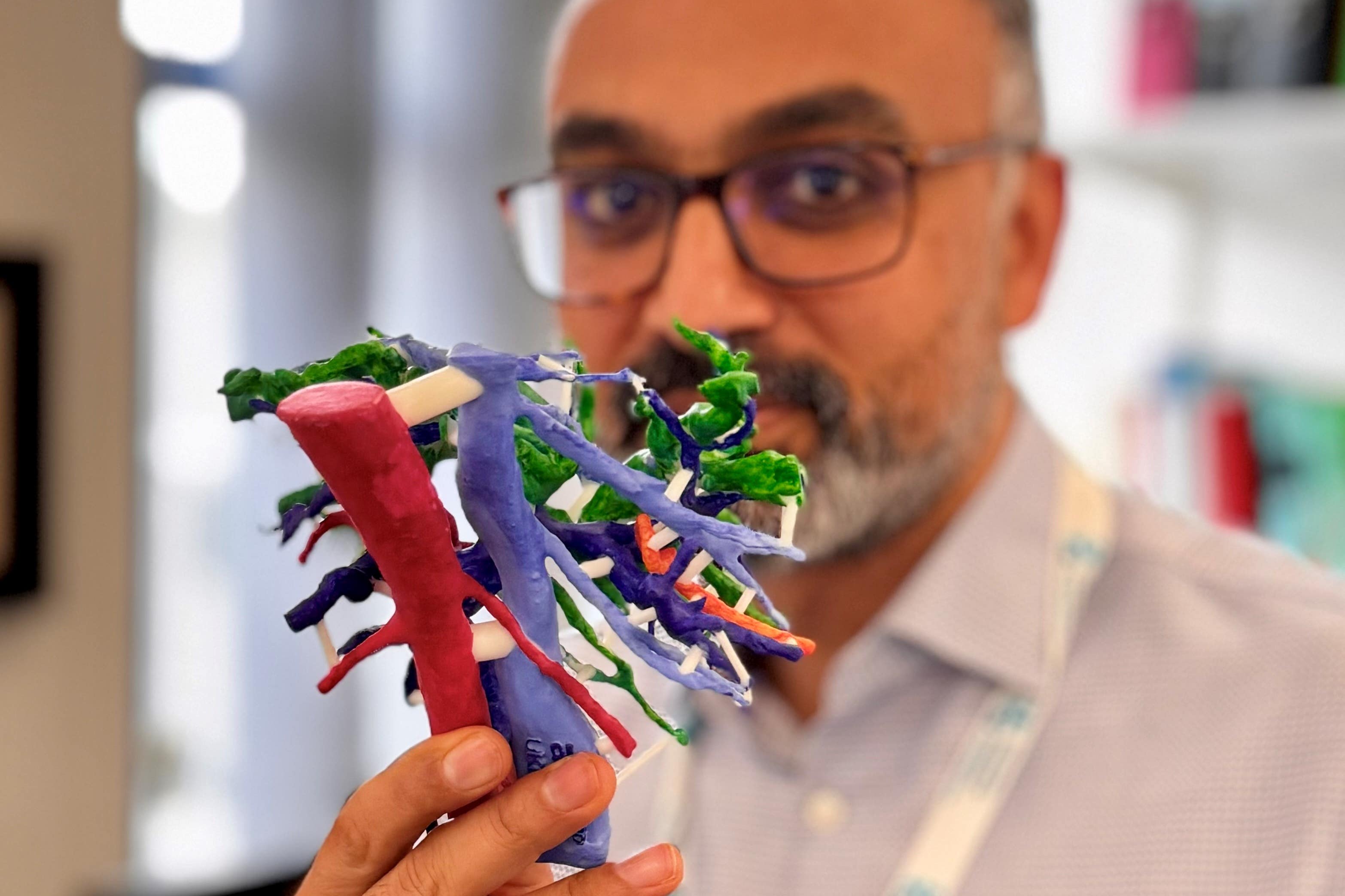
Surgeons develop 3D-printed liver models to plan complex cancer surgery
Data from CT and MRI scans of patients with hilar cholangiocarcinoma, a type of bile duct cancer, is used to create detailed physical models.
Your support helps us to tell the story
From reproductive rights to climate change to Big Tech, The Independent is on the ground when the story is developing. Whether it's investigating the financials of Elon Musk's pro-Trump PAC or producing our latest documentary, 'The A Word', which shines a light on the American women fighting for reproductive rights, we know how important it is to parse out the facts from the messaging.
At such a critical moment in US history, we need reporters on the ground. Your donation allows us to keep sending journalists to speak to both sides of the story.
The Independent is trusted by Americans across the entire political spectrum. And unlike many other quality news outlets, we choose not to lock Americans out of our reporting and analysis with paywalls. We believe quality journalism should be available to everyone, paid for by those who can afford it.
Your support makes all the difference.Surgeons have launched a new technique to use 3D printed models of patients’ livers to help them perform a complex cancer operation.
Medics at University Hospital Southampton have been supported by the Planets Cancer Charity to help produce the models, which are tailored to each patient to plan surgery and to ensure the best outcomes from “irreversible steps” carried out during an operation.
Data from CT and MRI scans of patients with hilar cholangiocarcinoma, a type of bile duct cancer, is used to create detailed physical models.
These are then analysed by the surgeons to establish whether a tumour can be safely removed, a decision that previously could not be made until the operation was actually under way.
Arjun Takhar, consultant hepatobiliary and pancreatic cancer surgeon, said: “3D printed models are increasingly being used to help with decision-making before and during surgery and to better understand the anatomical relationships of tumours within organ structures.
“With this particularly challenging liver/bile duct cancer, hilar cholangiocarcinoma, sometimes we are unable to tell until the very last minute whether the tumour can be safely removed or not.
“Sometimes surgeons will have taken an irreversible step and then found out that the tumour cannot be removed completely, which results in poor outcomes for patients in the short and long term.”
Planets Cancer Charity has received a £2,000 grant from The Hospital Saturday Fund for the pilot initiative.
Mr Takhar said: “3D printing offers the advantage of assessing the tumour and its close attachments such as blood vessels and bile ducts in a scale model prior to performing the operation itself.
“The aim of the pilot project is to assess whether this allows for assessment of operability to be made without opening the patient up, and also whether assessment with a model is better than simply looking at the patient’s CT and MRI scans.
“There is also an added advantage that a 3D model would also help in teaching trainee surgeons the nuances of liver anatomy in relation to these complex tumours.
“This is a unique opportunity to use a novel technology to help patients with a difficult disease and we foresee adoption of the technology in patients with other liver tumours too.”
Planets helps patients with pancreatic, liver, colorectal, abdominal (oesophageal and gastric) and neuroendocrine cancer by funding patient support groups, innovative treatments and research primarily in Hampshire, Wiltshire, Dorset, Isle of Wight, Channel Islands and West Sussex.